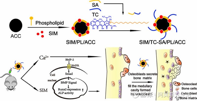
A biological mechanism by which calcium supplement using ACC augments Sim mediated BMP-2-induced osteoblast differentiation is shown as Runx2 expression and ALP activity through the BMP-Smad signaling pathway.【如图所示,ACC钙补充剂可促进BMP-2诱导间充质细胞定向分化为成骨细胞,通过成骨细胞碱性磷酸酶ALP活性调控BMP-Smad信号通路】
背景:
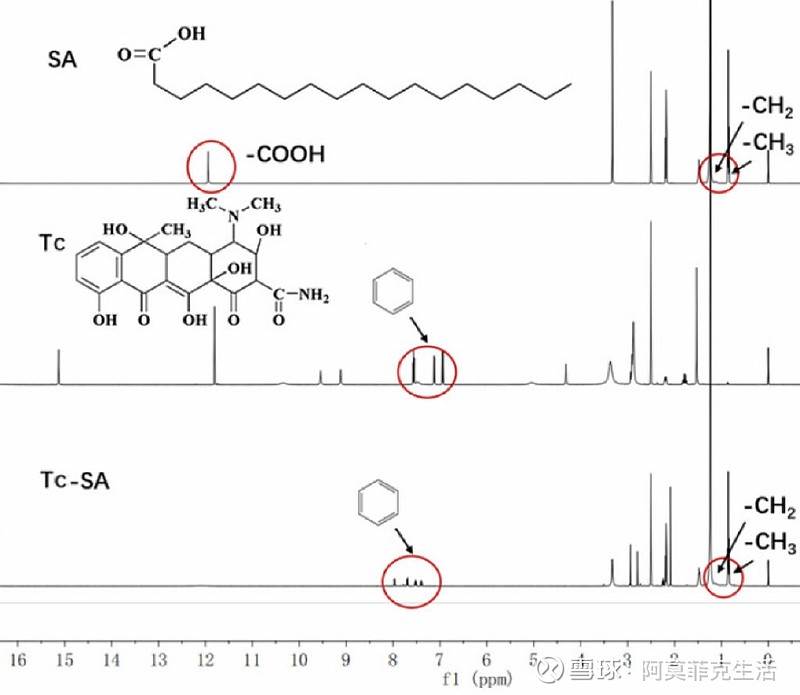
Characterization of Tc-SA using 1H NMR. Inserted images were corresponding chemical structure and peak assignments.【1H-NMR谱图解析Tc-SA的分子结构及各峰的位置(化学位移)】
钙补充剂是一种临床批准的骨质疏松症治疗方法,但通常需要大剂量且无靶向性且结果不佳。
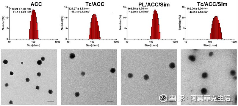
Characterization of Tc/ACC from ACC using (A) dynamic light scattering (DLS) and (B) transmission electron microscopy (TEM) images. Scale bar: 100 nm.【使用(A)动态光散射(DLS)和(B)透射电子显微镜(TEM)图像表征ACC的Tc / ACC。比例尺:100纳米。】
由于缺乏适当的钙补充剂载体,这种方式在目前的骨质疏松症治疗中没有得到充分探索。
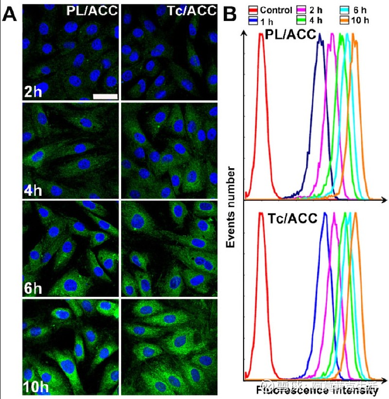
In vitro cellular uptake of Tc/ACC. MC3T3-E1 cells were incubated with ODA-FITC labeled PL/ACC and Tc/ACC for different time intervals (2, 4, 6, 10 h) and then subjected to observation using confocal microscope. (A) Time-dependent cellular uptake of ODA-FITC labeled PL/ACC and Tc/ACC. Scale bar: 30 µm. (B) Semiquantitative analysis of cellular uptake by flow cytometry.【MC3T3-E1细胞与ODA-FITC标记的PL / ACC和Tc / ACC一起孵育不同时间间隔(2,4,6,10 h)的体外细胞摄取,然后使用共聚焦显微镜进行观察。(A)ODA-FITC标记的PL / ACC和Tc / ACC的时间依赖性细胞摄取。规模条:30μm。】
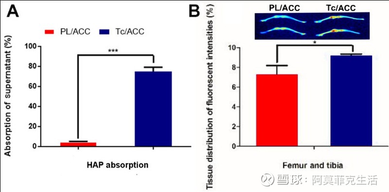
Bone targeting of Tc/ACC. (A) In vitro bone targeting of Tc/ACC using HAP adsorption assay. HAP was added to the ODA-FITC labeled PL/ACC and Tc/ACC and incubated for under genteel mechanical stirring for 1 hour. Afterwards, the decrease in the fluorescence of the supernatant was measured. (B) In vivo bone targeting of Tc/ACC using fluorescence imaging. ICR mice were randomly divided into two groups and intravenously injected with ICG labeled PL/ACC and Tc/ACC. At 48 h post administration, the mice were sacrificed and the quantitative accumulation of fluorescence signal in the femur, and tibia bones were calculated. Inserted image was fluorescence images of the distribution of ICG labeled PL/ACC and Tc/ACC in the femur and tibia bones. Results were expressed as mean ± S.D. (n = 3).【(A)使用HAP吸附测定法对Tc / ACC进行体外骨靶向。将HAP加入到标有PL / ACC和Tc / ACC的ODA-FITC中,并在温和的机械搅拌下孵育1小时。之后,测量上清液荧光的降低。(B)使用荧光成像在体内靶向Tc / ACC的骨。将ICR小鼠随机分为两组,并静脉注射ICG标记PL / ACC和Tc / ACC。在给药后48 h处死小鼠,计算股骨中荧光信号的定量积累,并计算胫骨。插入的图像是ICG在股骨和胫骨中分配的PL / ACC和Tc / ACC的荧光图像。结果表示为平均±S.D.(n = 3)。】
方法:
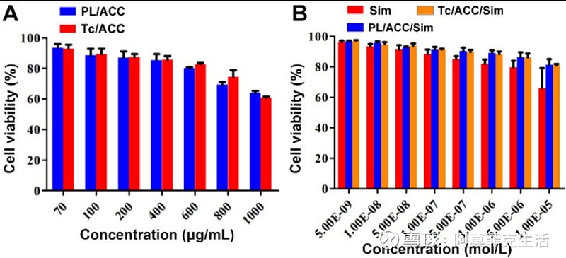
Optimization of administration dosage. MC3T3-E1 cells were exposed to a series of concentrations of PL/ACC, Tc/ACC, Sim, PL/ACC/Sim and Tc/ACC/Sim for 48 h. Afterwards, the cell viability influence of (A) Sim free carrier (PL/ACC and Tc/ACC) and (B) Sim containing formulations were evaluated using MTT assay. Results were expressed as mean ± S.D. (n = 3).【优化给药剂量。将MC3T3-E1细胞暴露于一系列浓度的PL / ACC,Tc / ACC,Sim,PL / ACC / Sim和Tc / ACC / Sim中48小时。之后,使用MTT测定法评估了(A)不含Sim的载体(PL / ACC和Tc / ACC)和(B)含Sim制剂的细胞活力影响。结果表示为平均±S.D.(n = 3)。】
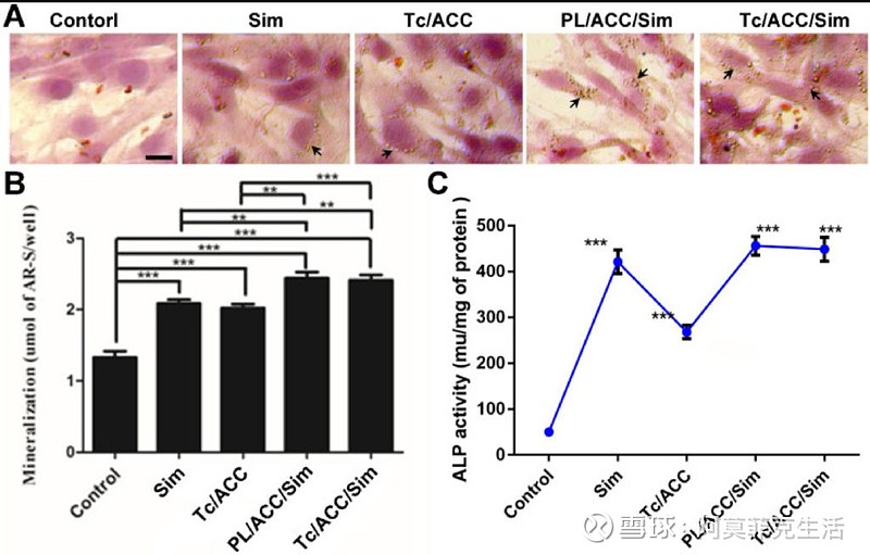
Effects of Sim-loaded nanoparticles on mineralized nodule formation and ALP activity in MC3T3-E1 cells. MC3T3-E1 cells were cultured in standard osteogenic differentiation and mineralization medium containing 10 mM β-glycerophosphate, 50 µg/mL ascorbic acid and different formulations at the equal Sim concentration of 10-7 M). Afterwards, the mineralization of the extracellular matrix was evaluated by optical microscopy and macroscopic observation (A), quantitative mineralization results (B) at 14 days post incubation, and stimulation of ALP activity (C) at 7 days post incubation. Results were expressed as mean ± S.D. (n = 3). *p < 0.05, **p < 0.01, ***p < 0.001, significant differences compared with the control.【Sim负载纳米颗粒对MC3T3-E1细胞矿化结核形成和ALP活性的影响.将MC3T3-E1细胞在含有10mM β-甘油磷酸,50μg/ mL抗坏血酸和不同制剂的标准成骨分化和矿化培养基中培养,Sim浓度相等,为10-7 M)。之后,通过光学显微镜和宏观观察(A)评估细胞外基质的矿化,孵育后14天定量矿化结果(B),孵育后7 d刺激ALP活性(C)。结果表示为平均±S.D.(n = 3)。*p < 0.05,**p < 0.01,***p < 0.001,与对照组相比差异显著。】
在本研究中,我们构建了四环素 (Tc) 修饰和辛伐他汀 (Sim) 负载的磷脂-无定形碳酸钙 (ACC) 杂化纳米颗粒 (Tc/ACC/Sim)。
结果:
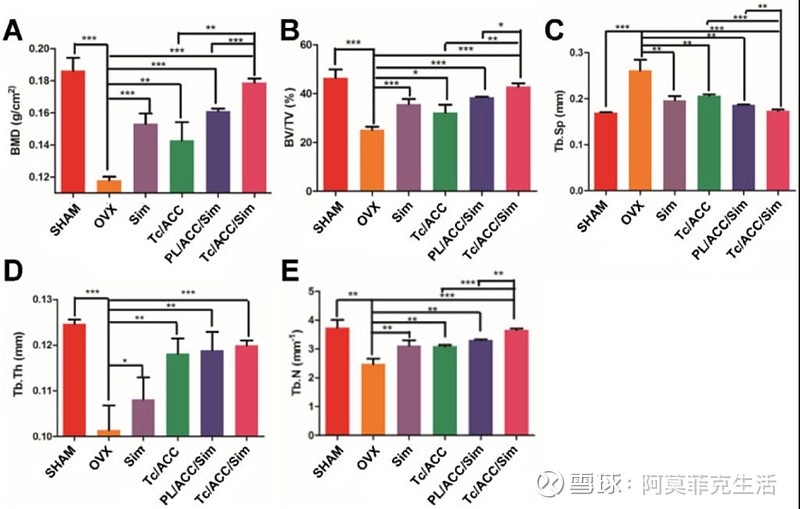
Female Sprague Dawley rats were randomly selected and subjected to sham operation or bilaterally oophorectomy. The treatment groups were intravenously administered with Sim, PL/ACC/Sim or Tc/ACC/Sim group at the Sim dose of 1 mg/kg/2 days for 2 months. Afterwards, the rats were sacrificed and the micro-CT analysis of bone tissue from each group were conducted. (A) Trabecular bone density (BMD); (B) Trabecular bone volume percentage (BV/TV); (C) Trabecular separation (Tb.Sp); (D) Trabecular thickness (Tb.Th) and (E) trabecular number (Tb.N) of the femur. Results were expressed as mean ± S.D. (n = 3). *p < 0.05, **p < 0.01, ***p < 0.001, significant differences compared with the control.【随机选择雌性Sprague Dawley大鼠并进行假手术或双侧卵巢切除术。治疗组与Sim,PL / ACC / Sim或Tc / ACC / Sim组以1mg / kg / 2天的Sim剂量静脉内给药,持续2个月。之后,处死大鼠,并对每组骨组织进行微CT分析。(A) 小梁骨密度(BMD);(B) 小梁骨体积百分比(BV/TV);(C) 小梁分离(Tb.Sp);(D)股骨的小梁厚度(Tb.Th)和(E)小梁数(Tb.N)。结果表示为平均±S.D.(n = 3)。*p < 0.05,**p < 0.01,***p < 0.001,与对照组相比差异显著。】
所得的 Tc/ACC/Sim 能够增强其在骨质疏松部位的积累。最重要的是,钙补充剂和 Sim 的组合提供了骨质疏松症的协同成骨细胞促进治疗,其性能优于非靶向系统或单一治疗。
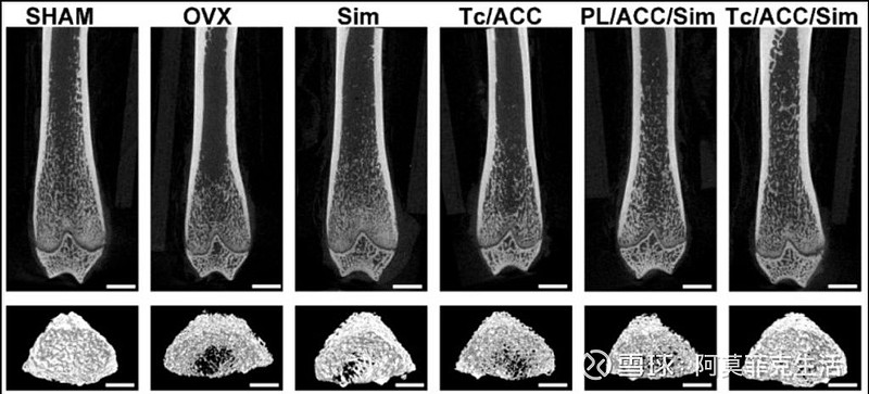
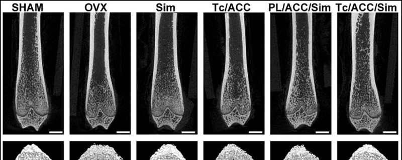
The 2D and 3D images of trabecular bone measured by micro-CT. Scale bar: 1mm.【通过显微CT测量的小梁骨的2D和3D图像。比例尺:1毫米。】
结论:
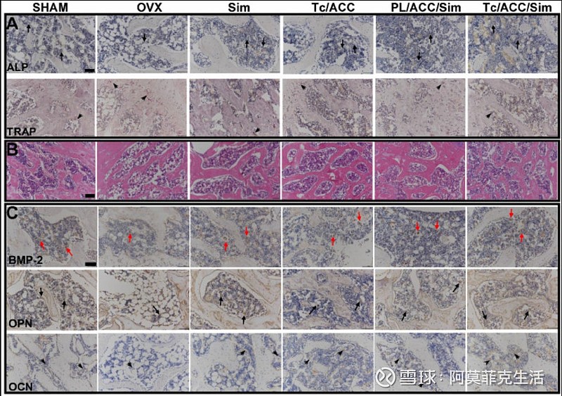
(A)ALP activity (arrows) and TRAP assay results (arrowheads) of bone tissue sections. (B) Histological assessment of bone formation in each group using HE staining. (C) Immunohistochemical staining for BMP-2 in typical newly-formed bone tissue (red arrows) and immunohistochemical staining for the osteogenic markers OPN (arrows) and OCN (arrowheads). Scale bars = 100 µm.【(一)骨组织切片的ALP活性(箭头)和TRAP测定结果(箭头)。(B)使用HE染色对每组骨形成进行组织学评估。(C)在典型的新形成的骨组织(红色箭头)中对BMP-2进行免疫组织化学染色,对成骨标志物OPN(箭头)和OCN(箭头)进行免疫组织化学染色。比例尺 = 100 μm。】
该平台提供了一种通过使用钙补充剂和 Sim 协同促进成骨细胞分化来刺激骨形成的替代方法。
原文链接:网页链接